Device-Based Management of Rhythm Disorders
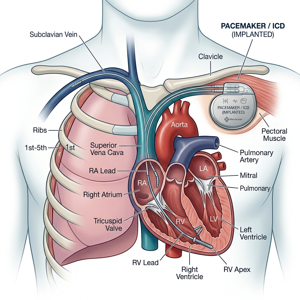
Cardiac pacemakers and implantable cardioverter-defibrillators (ICDs) are sophisticated electronic devices placed inside the body to monitor and correct disturbances in the heart's electrical system. A pacemaker is primarily indicated when the heart beats too slowly (bradycardia), stepping in to deliver electrical impulses that sustain a safe, regular heart rate. An ICD is a life-saving device designed to detect excessively fast and chaotic, life-threatening arrhythmias (such as ventricular tachycardia or fibrillation) within seconds, administering pacing therapies or an electrical shock to immediately restore a normal sinus rhythm and prevent sudden cardiac death.
Implantation is a minor surgical procedure, not open-heart surgery. Under local anesthesia, a small incision is made just below the collarbone to create a pocket under the skin. Thin, insulated wires (leads) are guided through a vein into the heart chambers and secured. The leads are then connected to the generator (the battery pack), which is placed in the subcutaneous pocket. Recovery is quick, and patients are typically discharged the next day to return to daily living shortly after.
Indications for Pacemakers and ICDs
- Patients with severe bradycardia due to heart blocks (AV blocks) or sick sinus syndrome experiencing dizziness, fatigue, or fainting (syncope).
- Heart failure patients requiring cardiac resynchronization therapy (CRT) using a specialized three-lead biventricular pacemaker to improve heart pumping efficiency.
- Patients with advanced heart failure and a reduced ejection fraction (EF ≤ 35%) after a heart attack who carry a high risk for sudden cardiac arrest (ICD recommendation).
- Survivors of a previous sudden cardiac arrest or individuals with genetic cardiac conditions predisposed to dangerous ventricular rhythms.
The Pacemaker Implantation Steps
-
01
Step 1
Evaluation & Device Selection
Diagnostic workup ( Holter EKG, echocardiography, etc.) is reviewed to decide the optimal device configuration (single, dual chamber pacemaker, ICD, or CRT).
-
02
Step 2
Lead Insertion
The area under the collarbone is numbed. Under fluoroscopic guidance, the thin insulated leads are advanced through a vein and positioned securely within the heart chambers.
-
03
Step 3
Generator Connection
A small pocket is created underneath the skin/fascia. The leads are connected to the generator body, and the generator is placed inside the pocket.
-
04
Step 4
Programming & Calibration
Electrical thresholds are verified. The device settings are programmed wirelessly to match the patient's individual heart rate and therapy requirements, and the incision is closed.
Benefits of Pacemaker & ICD Treatment
Academic Quality & Advanced Device Programming
- High volume of pacemaker, ICD, and CRT implants performed successfully at Gazi University.
- Academic coordination of routine follow-up, wireless programming, and threshold calibrations.
- Technical proficiency in placing biventricular leads in challenging coronary sinus networks.
- A patient-first, transparent standard of care focusing strictly on ethical device indications.
FAQ About Pacemakers & ICDs
A pacemaker regulates the rhythm when the heart beats too slowly. An ICD yavaş ritimleri düzenlemenin yanı sıra, ani gelişebilecek ölümcül hızlı ritimleri algılayıp şok vererek hayatı kurtarma yeteneğine sahiptir (defibrillator function).
During the first few weeks, avoid lifting heavy objects or reaching high above shoulder height with the arm on the device side. Walk normally through security metal detectors. Avoid placing cell phones directly over the device pocket. Most modern devices are MRI-compatible, though settings must be adjusted before any scan.
The battery lasts 6 to 10 years depending on the device type and utilization. Impending depletion is detected months in advance during routine checks. Battery replacement is a simple 20-30 minute procedure where the pocket is opened under local anesthesia, and the generator box is swapped out without touching the leads in the heart.
For the first 4-6 weeks, you should restrict large, sudden arm extensions or lifting heavy weights on the device side to allow the leads to anchor firmly in the heart tissue. Once this period has passed, the incision is fully healed, and there are no limitations on arm movement.